
Hygiene in veterinary theatres: what a clean-looking surface will not tell you
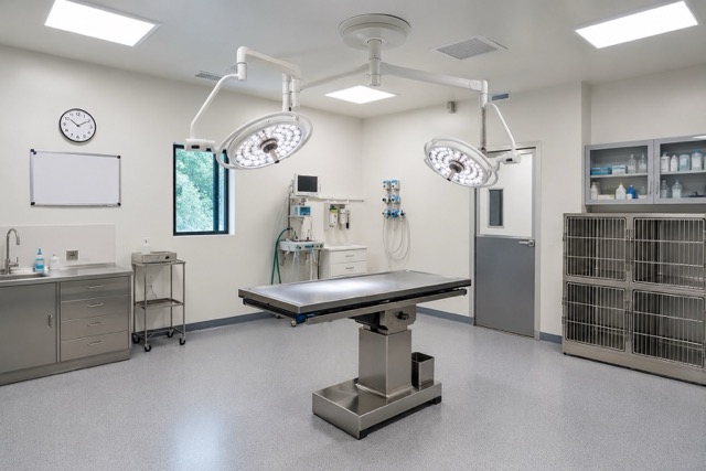
Walk into a well-run small animal theatre at eight o’clock in the morning and it looks immaculate. Surfaces wiped down, equipment laid out, floor still damp from the overnight clean, a practice which is peaceful and ready for their first patient.
 A lot of effort goes into creating a clean environment yet surgical site infection rates in clean small animal procedures sit roughly between 2.5% and 4.8% depending on the study and the procedure and some operations run higher. Tibial plateau levelling osteotomy has been associated with rates of around 7%. One study of 1,550 dogs recorded surgical site infections in 85 cases (5.5% overall) that were split fairly evenly between orthopaedic and soft tissue work being done.
Those are not catastrophic numbers but behind every percentage point sits a returning ill patient, a course of antibiotics that should not have been needed, an awkward conversation with an owner and a bill somebody has to absorb.
The risk is not where most people look
If you ask a practice team where infection risk lives, they usually point at the surgical field or the instrument tray. Both areas are heavily controlled however studies suggest the risks are elsewhere.
A study of 60 clipper blades in small animal private practice found 51% were contaminated with bacteria, most commonly Staphylococcus species and Coliforms. Surgical practices came off worse than dermatology practices, 67% against 13%. A more recent Swedish study went further: between 64% and 100% of disinfected blades across three hospitals still grew bacteria, while sterilised blades came back clean every time. Clippers touch patient skin minutes before incision is made which is a direct route of entry.
As well as the operating field there is the anaesthesia workspace. A six-month hygiene audit at a US veterinary medical centre using traditional ATP bioluminescence found anaesthesia carts and touchscreen monitors among the most frequently contaminated sites, largely because nobody had flagged them as high-risk on a cleaning rota. Separate research on small animal breathing systems has shown they pick up environmental organisms during routine use.
Prep sinks, drains, wet tables and hydrotherapy kit is where Pseudomonas aeruginosa survives by forming biofilm on stainless steel, plastic and glass, then tolerating the disinfectants aimed at it. Research on hospital sink drains found biofilms regrew to baseline density in roughly four days after disinfection and the regrown community carried more resistant organisms than it had before.
Biofilm is the crucial part disinfectant misses
Routine cleaning lifts visible soil and kills what is exposed. It does not reliably remove a mature biofilm and a partly disrupted biofilm can shed viable organisms back onto a surface that now looks perfectly clean.
One veterinary teaching hospital found Methicillin-resistant Staphylococci (MRSP) widely distributed and persistent across surfaces despite routine, extensive cleaning with detergent and disinfectant. Of 88 coagulase-positive isolates, 70.5% were Methicillin resistant and MRSP accounted for almost 92% of those. Work at other hospitals has turned up MRSA on doors and trolleys, surfaces handled by dozens of people and patients a day.
When this goes wrong at scale it gets expensive fast. In a survey of veterinary hospitals reporting a nosocomial outbreak, 71% restricted admissions and 38% closed part or all of the facility. One equine teaching hospital closure has cost at over $4 million. Another put losses above $2 million once lost revenue, renovation, deep cleaning and ten months of reduced caseload were added up.
Visual inspection is not verification
Most practices verify hygiene standards by looking at the environment. Visual inspection consistently overestimates cleanliness when checked against traditional ATP swabbing and microbiological culture and the gap widens on high-touch clinical surfaces.
ATP swabbing is a logical answer and plenty of practices already use it, but it normally isn’t conducted that frequently and does have its limitations. A swab tells you about the few square centimetres you swabbed. Across a theatre, prep room and recovery area you are sampling a fraction of a percent of the total surface and trusting it to be representative. Swab the wrong spot (or right?) and a contaminated room passes inspection.
Seeing the problem before you sample it-making the invisible, visible
EIT International’s Bactiscan™ uses wave-alternating UV light to reveal biofilms on surfaces in real time. No chemicals, no dyes, no culture wait and for most areas no downtime. One person walks a theatre with a handheld unit and sees where contamination actually is and this is a simple step to add into an established workflow.
The technology is already used in food, beverage and pharmaceutical manufacturing, where a failed hygiene audit stops a production line. The logic transfers easily to a veterinary environment. Scan first, find the hot spots, then use ATP swabs where they will tell you something useful. This process gets better data from fewer swabs, saving time, saving money and doing a lot less guesswork.
For a practice that means a few practical things. Cleaning protocols get validated against what is genuinely on the surface rather than what the SOP assumes. Problem areas that survive every clean, the under edges of prep sinks, the underside of a wet table lip, the clipper charging station, get identified and dealt with properly. The BactiscanPRO model captures stills and videos, so remedial work can be documented for RCVS Practice Standards evidence, an accreditation visit, or an insurer asking questions after an incident.
Worth a conversation
No device prevents infection on its own. Hand hygiene, sterilisation, antimicrobial stewardship and surgical technique still do the heavy lifting and always will.
What scanning the environment does is close one specific gap: the reality between a surface that looks clean and a surface that is clean. Given what the environmental data shows about how stubbornly these organisms persist in veterinary hospitals and what a single outbreak costs when it takes hold, that reality is worth measuring.
To arrange a demonstration of Bactiscan in your practice, get in touch.
References
SSI rates in small animal surgery, CABI Digital Library / JAVMA: https://www.cabidigitallibrary.org/doi/10.1079/9781789244977.0009
Risk factors for SSI in clean surgical procedures in dogs: https://pubmed.ncbi.nlm.nih.gov/33905955/
Bacterial contamination of clipper blades in small animal private practice (JAAHA): https://jaaha.kglmeridian.com/view/journals/aaha/52/2/article-p95.xml
Microbes on clipper blades after use and disinfection: https://pubmed.ncbi.nlm.nih.gov/38250944/
ATP bioluminescence for hygiene assessment in a veterinary medical center (AJVR): https://avmajournals.avma.org/view/journals/ajvr/86/6/ajvr.24.09.0278.xml
ATP bioluminescence for monitoring surface hygiene in hospital settings (review): https://link.springer.com/article/10.1186/s13756-026-01698-8
Bacterial contamination of small animal breathing systems during routine use: https://pubmed.ncbi.nlm.nih.gov/17444932/
Veterinary clinics as reservoirs for Pseudomonas aeruginosa (Antibiotics): https://www.mdpi.com/2079-6382/14/7/720
Disinfection of hospital sink drains and biofilm regrowth (Nature Communications): https://www.nature.com/articles/s41467-026-73533-y
Methicillin-resistant coagulase-positive staphylococci in a veterinary teaching hospital: https://veterinaryworld.org/Vol.18/December-2025/3.php
Environmental MRSA in a veterinary teaching hospital: https://pmc.ncbi.nlm.nih.gov/articles/PMC3391706/
Environmental microbiological surveillance in veterinary hospitals (JAC-AMR): https://academic.oup.com/jacamr/article/6/4/dlae113/7721433
Barrier precautions and outbreak impact in veterinary hospitals: https://pmc.ncbi.nlm.nih.gov/articles/PMC7135499/
Salmonella Newport outbreak in a large animal veterinary teaching hospital: https://pubmed.ncbi.nlm.nih.gov/20584143/